Ultrasound’ Semiology Semiología de los Ultrasonidos
Sonography is a scanning technique inside the body using ultrasounds, which records the reflections or echoes in its propagation by internal discontinuities. Because of different organ and tissues acoustic impedance, and because of interface between them, we can get an image.
According to amount of echoes reflected by tissues we will get different images and we can classify organs and tissues like:
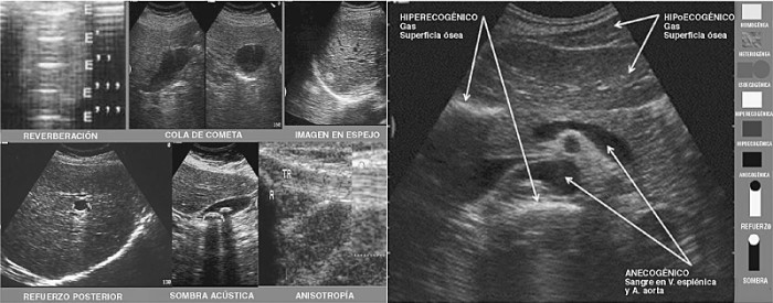
Echoic or echogenic: it is said about a tissue or structure that has or issue echoes. It is showed in a large greyscale depending on the characteristics of the reflected echo. Solid organs use to be echogenic. Depending on the echoes intensity, tissues will be:
- Hiperechoic or hiperechogenic: the transducer receives great quantity of echoes; therefore the structure is more echogenic than the rest of tissues that surround it.
- Isoechoic or isoechogenic: the transducer receives the same quantity of echoes of a structure that of other one. It is said of two structures with the same echogenicity, as the structure on which we centre as the tissue that surrounds it.
- Hypoechoic or hypoechogenic: the transducer receives less quantity of echoes; therefore the structure has less echogenicity than the rest of tissues that surround it.
Anechoic or anechogenic: in these structures the transducer doesn´t receive any echo, the structure doesn`t have any echo inside. The liquids, as the urine in the bladder, the bile in the gallbladder or the blood in the vessels; are anechoic and we see them black. Solid very homogeneous structures also might be anechoic.
Behind of the different tissues or structures that we visualize in an ultrasound image, we can find several appliances. The most common are:
- Posterior acoustic enhancement: it takes place when the ultrasound beam crosses a structure that attenuates a little bit the sound, so deeper structure or tissues will be seen by major echogenicity. This acoustic enhancement takes place after structures full of liquid, since the faeces aren´t reflected or greatly attenuate. (1)
- Sonic shade: it takes place when the ultrasound beam hits against an interface and crosses little or no sound across her. This brings as result that the whole beam is reflected, and below this one absence of sign is observed. The shade can be clean, if it is totally anechoic; and dirty, if some echoes are gathered. (1)
- Reverberation: it takes place when the echoes that are reflected and gathered by the transducer return to enter the patient again. This would produce the second echo that in the image will appear to the double of the distance of the first echo or real echo. This process can repeat successively and in the image will appear hyperechogenic parallel lines that decrease the intensity with increasing attenuation and depth. (2) It takes place in interfaces of tissue with very different acoustic impedance.
- Appliance in mirror or image to speculate: It takes place when the ultrasound beam has an impact on a curvilinear structure. Some echoes can change his path and bounce against another interface that reflects them towards the transducer (they come later so they do more travel). Physiological example: diaphragm. Part of the liver is reflected to another side of the diaphragm when it is known that to another side the air of the lung is. Pathological example: tumour next the diaphragm. (3)
- Comet tail: it takes place when the ultrasound beam hits against a not very thick interface and very echogenic, appearing behind this interface a series of linear echoes. Gases use to give images in tail of comet.
Bibliography
- Solano A C, Bernal G A, Espinosa M R, Hernández D C, Marín A N, Peña A A, et al. Artefactos en ecografía musculoesquelética. Revista chilena de reumatología. 2009; 25(2): p. 76-81.
https://www.sochire.cl/bases/r-386-1-1343744086.pdf
- Díez Bru N. Artículo de revisión. Principios básicos de la ecografía. Clinica veterinaria de pequeños animales. 1992 Julio/Septiembre; 12(3).
https://ddd.uab.cat/pub/clivetpeqani/11307064v12n3/11307064v12n3p138.pdf
- Díaz Rodríguez N, Garrido Chamorro R, Castellano Alarcón J. Metodología y técnicas. Ecografía: principios físicos, ecógrafos y lenguaje ecográfico. SEMERGEN-Medicina de familia. 2007 Agosto; 33(7).
Notes of the subject medical Image and Instrumentation. Course 2014/2015. Lesson 2: Ultrasounds. Technical and topographic bases of the medical ultrasound scan.
Author: Fátima Cabezas Fernández
2º Course, Medicine. Granada University
La ecografía es una técnica de exploración del interior de un cuerpo mediante ultrasonidos, que registra las reflexiones o ecos producidos en su propagación por las discontinuidades internas. Gracias a la diferente impedancia acústica de órganos y tejidos, y la existencia de interfase entre ellos podremos obtener una imagen.
Según la cantidad de ecos que reflejen los tejidos obtendremos diferentes imágenes y podremos clasificar las diversas estructuras en:
Ecoico o Ecogénico: se dice de aquel tejido o estructura que tiene o emites ecos. Se representa en una amplia escala de grises en función de las características del eco reflejado. Suelen ser órganos sólidos. En función de la intensidad de estos ecos los tejidos serán:
- Hiperecoico o Hiperecogénico: el transductor recibe gran cantidad de ecos, por tanto la estructura posee una mayor ecogenicidad que el resto de tejidos que la rodean.
- Isoecoico o Isoecogénico: el transductor recibe la misma cantidad de ecos de una estructura que de otra. Se dice de dos estructuras que tiene la misma ecogenicidad, tanto la estructura en la que nos centramos como el tejido que la rodea.
- Hipoecoico o Hipoecogénico: el transductor recibe menor cantidad de ecos, por tanto la estructura posee una menor ecogenicidad que el resto de tejidos que la rodean.
Anecoico o Anecogénico: en estas estructuras el transductor no recibe ningún eco, la estructura no tiene ningún eco en su interior. Se ve de color negro. Los líquidos, como la orina en la vejiga, la bilis en la vesícula o la sangre en los vasos; son anecoicos. También podrían ser anecoicas estructuras sólidas muy homogéneas.
Tras los diferentes tejidos o estructuras que visualizamos en una imagen ecográfica podemos encontrar diversos artefactos. Los más comunes son:
- Refuerzo sónico posterior: se produce cuando el haz ultrasónico atraviesa una estructura que atenúa poco el sonido, de manera que estructuras o tejidos más profundos se verán con mayor ecogenicidad. Este refuerzo sónico se produce tras estructuras llenas de líquido, pues los haces no se reflejan ni se atenúan en gran medida. (1)
- Sombra sónica: se producen cuando el haz ultrasónico choca contra una interfase y pasa poco o ningún sonido a través de ella. Esto trae como resultado que todo el haz sea reflejado, y por debajo de éste se observe ausencia de señal. La sombra puede ser limpia, si es totalmente anecoica; y sucia si se recogen algunos ecos. (1)
- Reverberación: se produce cuando los ecos que son reflejados y recogidos por el transductor y vuelven a entrar en el paciente de nuevo. Esto produciría un segundo eco que en la imagen aparecerá al doble de la distancia del primer eco o eco real. Este proceso puede repetirse sucesivamente y en la imagen aparecerán líneas hiperecogénicas paralelas que van disminuyendo de intensidad a medida que aumenta la atenuación y la profundidad. (2) Se produce en interfases de tejido con muy diferente impedancia acústica.
- Artefacto en espejo o imagen especular: Se produce cuando el haz de ultrasonidos incide sobre una estructura curvilínea. Algunos ecos pueden cambian su trayectoria y rebotan contra otra interfase que los refleje hacia el transductor (llegan más tarde pues hacen mayor recorrido).Ejemplo fisiológico: diafragma. Parte del hígado se ve reflejada al otro lado del diafragma cuando se sabe que al otro lado está el aire del pulmón. Ejemplo patológico: tumor próximo al diafragma. (3)
- Cola de cometa: se produce cuando el haz de ultrasonidos choca contra una interfase no muy gruesa y muy ecogénica, apareciendo detrás de esta interfase una serie de ecos lineales. Los gases suelen dar imágenes en cola de cometa.
Bibliografía
- Solano A C, Bernal G A, Espinosa M R, Hernández D C, Marín A N, Peña A A, et al. Artefactos en ecografía musculoesquelética. Revista chilena de reumatología. 2009; 25(2): p. 76-81.
https://www.sochire.cl/bases/r-386-1-1343744086.pdf
- Díez Bru N. Artículo de revisión. Principios básicos de la ecografía. Clinica veterinaria de pequeños animales. 1992 Julio/Septiembre; 12(3).
https://ddd.uab.cat/pub/clivetpeqani/11307064v12n3/11307064v12n3p138.pdf
- Díaz Rodríguez N, Garrido Chamorro R, Castellano Alarcón J. Metodología y técnicas. Ecografía: principios físicos, ecógrafos y lenguaje ecográfico. SEMERGEN-Medicina de familia. 2007 Agosto; 33(7).
Apuntes de la asignatura Imagen médica e Instrumentación. Curso 2014/2015. Lección 2: Ultrasonidos. Bases técnicas y topográficas de la ecografía médica.
Autora: Fátima Cabezas Fernández
2º Curso, Medicina. Universidad de Granada.


