Image Generation by Positron Emission Tomography (PET) Formación de Imágenes por Tomografía por Emisión de Positrones (PET)
The development of nuclear medicine has resulted in a massive application of effective routine imaging methods that have reached widely recognized clinical usefulness for the diagnosis and characterization of different dissorders1.
Positron Emission Tomography (PET) is one of the image techniques included in the above mencioned nuclear medicine. This consists of the introduction of a substance (radionuclide) inside the body, which requires having a short physical and biological period to irradiate the pacient the less possible. Besides, this radionuclide must generate enough radiation to be detected.
When radionuclides disintegrate, they turn into other radionuclides after emitting radiation (gamma, alpha, beta+ or beta-).
PET uses radionuclides that can emit “positrons” (beta-positive particles). These particles are very unstable as they quickly react against near beta-negative particles, producing the “annihilation reaction”.
Both the energy and mass of these two particles (beta+ and beta-) lead to the emission of two fotons with the same energy that propagate forming a 180º angle between them2. This property is used to form the image, as the patient is surrounded by several receptors that detect the coincident fotons generated by the positrons annihilation reactions. In this way, two 180º-angled fotons are detected simultaneously in the receptors ring.
Electronics for PET detectors can count the fotons and create fully three-dimensional images in few minutes, reducing clinical imaging times while improving imaging quality, with excellent space resolution and sensitivity3.
The radioisotopes used in PET are 11C, 15O, 13N and 18F. These atoms are very common in biomolecules and that’s why human body incorporates them easily. Moreover, their lifetime is quite short.
PET is essentially used in oncology, cardiology and neuropsychiatry, although it is also applied in many other domains. The problem of this technique is its price, provided that radionuclides have a very short lifetime, and thus they have to be generated continuously by a cyclotron, which is extremely expensive.
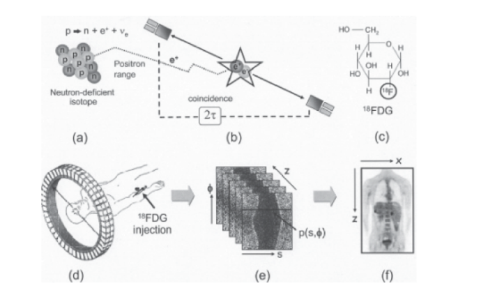
Figure 1: In the figure3 the steps that secuencially explain the image generation by PET can be observed.
Source: Townsend DW. Physical principles and technology of clinical PET imaging. Ann Acad Med Singapore. 2004 Mar;33(2):133-45.
REFERENCES
1. Ali Gholamrezanejhad, Sahar Mirpour, Giuliano Mariani. Future of Nuclear Medicine: SPECT Versus PET. J Nucl Med. 2009;50:16N-18N. Disponible en: https://jnm.snmjournals.org/content/50/7/16N.citation
2. Disselhorst JA, Bezrukov I, Kolb A, Parl C, Pichler BJ. Principles of PET/MR Imaging. J Nucl Med. 2014 May 12;55(Supplement 2):2S-10S. Disponible en: https://jnm.snmjournals.org/content/55/Supplement_2/2S.full.pdf+html
3. Townsend DW. Physical principles and technology of clinical PET imaging. Ann Acad Med Singapore. 2004 Mar; 33(2):133-45. Disponible en: https://www.annals.edu.sg/pdf200403/V33N2p133.pdf
Author: Mario Rivera Izquierdo
2º Course, Medicine. Granada University
El desarrollo de la medicina nuclear ha dado lugar a la aplicación a gran escala, de manera rutinaria, de métodos de imagen efectivos que han sido sumamente reconocidos por su utilidad clínica en el diagnóstico y caracterización de diferentes enfermedades1.
La tomografía por emisión de positrones (PET) forma parte de las técnicas de imagen que se engloban dentro de la ya citada «medicina nuclear». Esta consiste en la introducción de una sustancia (radionúclido) dentro del organismo, la cual debe poseer un período físico y biológico corto para irradiar de forma mínima al paciente pero al mismo tiempo debe generar una cantidad de radiación suficiente para permitir su adecuada detección.
Cuando los radionúclidos se desintegran, pasan a convertirse en otros radionúclidos tras emitir un tipo de radiación (que puede ser gamma, alfa, beta+ o beta-).
En el PET se emplean radionúclidos que son capaces de emitir positrones (partículas beta positivas). Estas partículas son muy inestables, al reaccionar con partículas beta negativas cercanas, produciéndose así la llamada «reacción de aniquilación».
La energía y masa de estas dos partículas (beta+ y beta-) se transforma dando lugar a dos fotones con la misma energía que se propagan formando un ángulo de 180º entre sí2. Esta propiedad es la que se utiliza para formar la imagen, pues alrededor del paciente se colocan diversos receptores capaces de detectar ambos fotones coincidentes formados en la reacción de aniquilación del positrón. De esta manera, en el anillo de receptores se van a detectar de forma simultánea dos fotones que forman un ángulo de 180º entre sí.
Los detectores electrónicos del PET son capaces de realizar el recuento de los fotones y generar una imagen tridimensional en minutos, con gran resolución espacial y alta sensibilidad3.
Los radioisótopos utilizados en el PET son 11C, 15O, 13N y 18F. Estos átomos son muy frecuentes en las biomoléculas, por lo que el cuerpo humano los incorpora con facilidad. Además, su vida media es muy corta.
El PET se utiliza fundamentalmente en oncología, cardiología, y neuropsiquiatría, aunque sus aplicaciones abarcan muchos otros ámbitos. El problema de esta técnica es su alto coste, dado que los radionúclidos tienen una vida media muy corta, por lo que es necesario generarlos mediante un ciclotrón (que es sumamente caro).
Figura 1: En la figura podemos observar los pasos que explican secuencialmente la formación de imágenes por PET.
Fuente: Townsend DW. Physical principles and technology of clinical PET imaging. Ann Acad Med Singapore. 2004 Mar;33(2):133-45.BIBLIOGRAFÍA
1. Ali Gholamrezanejhad, Sahar Mirpour, Giuliano Mariani. Future of Nuclear Medicine: SPECT Versus PET. J Nucl Med. 2009;50:16N-18N. Disponible en: https://jnm.snmjournals.org/content/50/7/16N.citation
2. Disselhorst JA, Bezrukov I, Kolb A, Parl C, Pichler BJ. Principles of PET/MR Imaging. J Nucl Med. 2014 May 12;55(Supplement 2):2S-10S. Disponible en: https://jnm.snmjournals.org/content/55/Supplement_2/2S.full.pdf+html
3. Townsend DW. Physical principles and technology of clinical PET imaging. Ann Acad Med Singapore. 2004 Mar;33(2):133-45. Disponible en: https://www.annals.edu.sg/pdf200403/V33N2p133.pdf
Autor: Mario Rivera Izquierdo
2º Curso, Medicina. Universidad de Granada.


